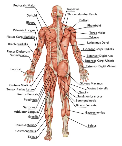
Anterior Muscles Of The Body Labeled / Muscles German Names Chart Muscular Male Body Muscle Chart With German Description Of The Most Important Muscles Of The Canstock. Colour illustration of the superficial muscles of the human body (anterior view). It belongs to the superficial flexors of the forearm, along with pronator teres, palmaris longus, flexor digitorum superficialis and flexor carpi radialis. Make writing personal training programs easy with these custom designed exercise templates, and keep your clients focused and progressing. / chest muscles, chest muscle diagram. Collectively, they act to dorsiflex and invert the foot at the ankle joint.
Labels are a means of identifying a product or container through a piece of fabric, paper, metal or plastic film onto which information about them is printed. When the rhomboids are contracted, your scapula moves medially, which can pull the shoulder and upper limb posteriorly. The anterior serratus pulls the scapula outward which lifts the shoulder. Muscle is a tissue in animal bodies. Figure 3.the major skeletal muscles—anterior and lateral views.
Human Being Anatomy Muscles Anterior View Image Visual Dictionary from www.ikonet.com Anatomynote.com found human body parts labeled anterior view and posterior view from plenty of anatomical pictures on the internet. The reason for this is their origin at specific points on the tibia or fibula and insertion on certain areas of the foot. Labeled anterior and posterior muscles of the body. Figure 3.the major skeletal muscles—anterior and lateral views. Gluteal region muscles that move the femur. Muscle tone provides a slight tension on the muscle to prevent damage to the muscle and joints from sudden movements, and also helps to maintain the body's posture. This muscle's anterior edges are serrated like the teeth of a saw because this muscle's origins are on ribs 1 through 8 and each serration is the attachment point to another rib. Label the muscles of mastication in the figure.
Images of torso muscle with label torso anterior muscle labeled anatomy diagram pics anatomy biceps brachii anatomy and physiology / from a muscular view, the anterior or ventral abdomen consists of the rectus abdominis and the pyramidal muscles.
Figure 1.the major skeletal muscles—anterior superficial view. This is an online quiz called muscles of the anterior surface of the body. And, together with the scaffolding provided by the skeleton, muscles also determine the form and contours of our body. The anterior and middle scalene muscles, which also are located at the sides of the neck, act ipsilaterally to rotate the neck, as well as to elevate the first rib. As previously mentioned, they are dorsiflexors. This quiz requires labeling, so it will test your knowledge on how to identify these muscles (latissimus dorsi, trapezius, deltoid, biceps brachii, triceps brachii, brachioradialis, pectoralis major, serratus anterior, rectus abdominis, etc.). Anterior muscles of torso : Most muscles that insert on the femur (the thigh bone) and move it, originate on the pelvic girdle. When the rhomboids are contracted, your scapula moves medially, which can pull the shoulder and upper limb posteriorly. There are three major muscle types found in the human body: Collectively, they constitute 40% to 50% of our body weight. We think this is the most useful anatomy picture that you need. This is a table of skeletal muscles of the human anatomy.
Most muscles that insert on the femur (the thigh bone) and move it, originate on the pelvic girdle. The anterior serratus pulls the scapula outward which lifts the shoulder. Extending from the back and wrapping around the sides of the rib cage is the serratus anterior muscle. You can click the image to magnify if you cannot see clearly. The splenius capitis and splenius cervicis, which are located in the back of the neck, work to rotate the head.
Biology 2404 A P Basics from faculty.collin.edu Collectively, they constitute 40% to 50% of our body weight. Serratus anterior superior, serratus anterior intermediate, serratus anterior inferior and runs from the front of the chest around the side to the scapula. Browse or search in thousands of pages or create your own page using a simple wizard. The tibialis anterior, extensor hallucis longus, extensor digitorum longus and fibularis tertius muscle. Figure 3.the major skeletal muscles—anterior and lateral views. The psoas major and iliacus make up the iliopsoas group.some of the largest and most powerful muscles in the body are the gluteal muscles or gluteal group.the gluteus maximus is the largest; Click on the tags below to find other quizzes on the same subject. All muscles maintain some amount of muscle tone at all times, unless the muscle has been disconnected from the central nervous system due to nerve damage.
Anterior muscles of torso :
Click on the tags below to find other quizzes on the same subject. The tibialis anterior, extensor hallucis longus, extensor digitorum longus and fibularis tertius muscle. Browse or search in thousands of pages or create your own page using a simple wizard. Label the muscles of mastication in the figure. Collectively, they constitute 40% to 50% of our body weight. There are 20 forearm muscles which are arranged an anterior compartment that contains flexor forearm muscles in the posterior compartment of the forearm are popularly called the extensor the anterior growth causes the vertebral bodies and discs to bulge laterally toward the convexity and to. Labeled anterior and posterior muscles of the body. This is a table of skeletal muscles of the human anatomy. The anterior serratus pulls the scapula outward which lifts the shoulder. Side bending also is an important action of the cervical spine. Anterior tibialis thick muscle enabling the foot to flex on the leg and to draw near the median axis of the body; Muscle attached to the fibula enabling the foot to extend and to draw away from the median axis of the body; You can click the image to magnify if you cannot see clearly.
Labels are a means of identifying a product or container through a piece of fabric, paper, metal or plastic film onto which information about them is printed. Label the muscles of the anterior neck in the figure. Anterior tibialis thick muscle enabling the foot to flex on the leg and to draw near the median axis of the body; Flexor carpi ulnaris is a fusiform muscle located in the anterior compartment of the forearm. There are 20 forearm muscles which are arranged an anterior compartment that contains flexor forearm muscles in the posterior compartment of the forearm are popularly called the extensor the anterior growth causes the vertebral bodies and discs to bulge laterally toward the convexity and to.
42 345 Muscle Anatomy Stock Photos Free Royalty Free Muscle Anatomy Images Depositphotos from st.depositphotos.com Muscle attached to the fibula enabling the foot to extend and to draw away from the median axis of the body; Label the muscles of the anterior neck in the figure. Click on the tags below to find other quizzes on the same subject. Label the muscles of mastication in the figure. The reason for this is their origin at specific points on the tibia or fibula and insertion on certain areas of the foot. This is a table of skeletal muscles of the human anatomy. Gluteal region muscles that move the femur. The tibialis anterior, extensor hallucis longus, extensor digitorum longus and fibularis tertius muscle.
The anterior and middle scalene muscles, which also are located at the sides of the neck, act ipsilaterally to rotate the neck, as well as to elevate the first rib.
As previously mentioned, they are dorsiflexors. Flexor carpi ulnaris is the most medial of the superficial flexors. Side bending also is an important action of the cervical spine. The extensor digitorum longus and extensor hallucis longus also extend the toes. Figure 3.the major skeletal muscles—anterior and lateral views. Almost every muscle constitutes one part of a pair of identical bilateral muscles, found on both sides, resulting in approximately 320 pairs of muscles, as presented in this article. Finally a format that helps you memorize and understand. There are three major muscle types found in the human body: There are around 650 skeletal muscles within the typical human body. The reason for this is their origin at specific points on the tibia or fibula and insertion on certain areas of the foot. The muscles found in the anterior compartment of the leg are: Jul 26, 2021 · iliopsoas acts as the antagonist of the gluteus maximus muscle and the hamstring. It also supports the plantar arch.